Obestetric Sonography - 4D/3D


Primary 3D or 4D ultrasound procedures, provide clearer insights into the developing fetus than conventional ultrasound. ّIt is done via comprehensive fetal anatomy screens and first-trimester fetal growth scans, such as moving images.
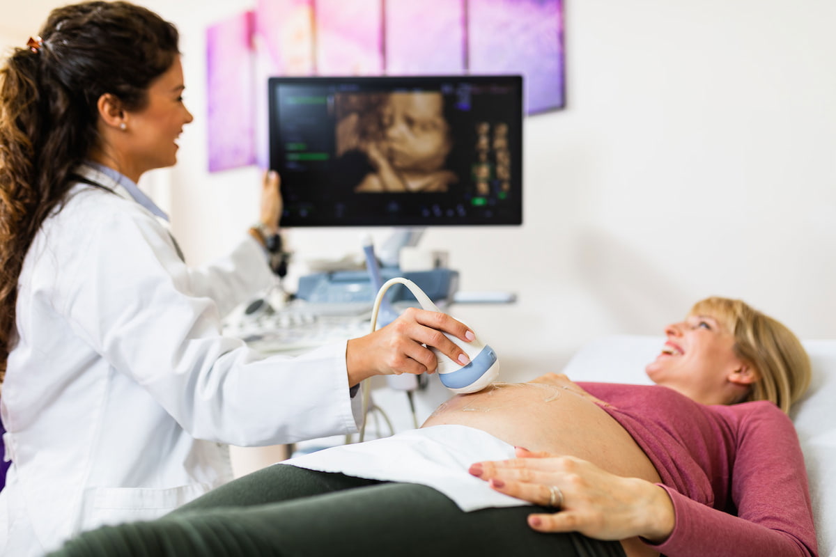
One of the modern technologies and therapeutic methods used to visualize the body’s internal parts is ultrasound, also known as diagnostic medical sonography, which is an imaging method for detecting and diagnosing a variety of diseases and conditions with the aim of directing them to proper treatment.
At Tadawi Medical Center, we offer you ultrasound imaging and obstetric ultrasound to help patients and pregnant women detect the internal organs of the body and assess their health condition under the supervision of expert doctors.
What is an ultrasound examination?
Ultrasound imaging is a highly accurate and easy-to-use medical imaging test. The device sends a group of high-frequency ultrasound waves to examine what is inside the abdomen, image fetuses inside the womb, detect aneurysms, and diagnose many health conditions, such as heart diseases and problems with the thyroid gland.
What is the purpose of obstetric ultrasound?
Obstetric ultrasound is one of the safe routine procedures that a pregnant woman resorts to since the first months of pregnancy for several reasons, namely:
- Examination of the uterus and ovaries in women.
- Check for defects or congenital problems in the fetus.
- Knowing the gestational age and ensuring the normal growth of the child.
- Determine the sex of the fetus and identify the number of fetuses in the womb.
What are the types of obstetric ultrasounds?
- Standard ultrasound
It is used to create two-dimensional images on a computer screen. It lasts about twenty minutes and gives information about the gestational age and assessment of the fetus.
- Ultrasound “Doppler”
This test measures the movement of blood flow and the speed at which blood travels through the uterus, umbilical cord, in the baby’s heart, or around the baby’s body.
- Fetal echocardiography
Designed to provide a more detailed picture of the chambers of the fetal heart and to check for or rule out congenital heart defects in the fetus.
- 3D – 4D ultrasound
The most advanced types of ultrasounds as providing a three or four-dimensional image of the fetus and detects congenital problems.
- Transvaginal ultrasound
One of the tests conducted in the first trimester of pregnancy gives a clearer picture of the fetus and its surrounding structures.
What month is best for an ultrasound scan?
An obstetric scan in pregnancy is performed at any time, but it is preferable to perform it between the sixth and twelfth week of pregnancy.
To detect and measure nuchal thickness in some serious pregnancies, the examination is repeated throughout the pregnancy to monitor the mother and fetus’s health carefully.
Does obstetric ultrasound affect pregnancy?
Obstetric ultrasonography is considered safe for the mother and the fetus’s health, as it does not use harmful rays, unlike x-rays or CT scans. Still, it is not advisable to overuse obstetric ultrasonography in anticipation of damage to the area exposed to radiation.
What is the difference between 3D and 4D ultrasound?
- 3D Ultrasound:
One of the techniques used is to photograph the interior of a woman’s uterus and display images in three dimensions on the screen, similar to three-dimensional films, but in a fixed and stereoscopic image so that the mother can see the fetus’s length and width.
- 4D Ultrasound:
One of the latest technologies used in obstetric ultrasonography, it displays a stereoscopic and integrated image like a 3D ultrasound. Still, its image is moving and more accurate so that the doctor can know more details about the fetus.
What can 3D or 4D ultrasound detect?
3D ultrasound is used for:
- Follow up on the pregnancy stages and ensure the fetus’s health and safety.
- Determining the sex of the fetus and the features of its shape more accurately than a regular ultrasound.
- Easily diagnose birth defects and hereditary or genetic diseases.
- Determine the location of the pregnancy sac inside or outside the uterus and the location of the umbilical cord.
- Ensure the growth and safety of the heart and internal organs of the embryos accurately.
- Study of the fluid around the fetus and blood nutrition of the organs.
4D ultrasound pregnancy is used for:
- Check on the health, size, and age of the fetus.
- Assessment of the child’s sleep, wakefulness, and complete movement of his organs.
- Early detection of some serious congenital anomalies.
- Early preparation for childbirth to save a child suffering from diseases.
- Detection of facial anomalies and the rabbit’s opening in the palate and lip.
Does 4D or 3D ultrasound detect fetal abnormalities?
Obstetric scans using 3D or 4D ultrasound enable parents and the doctor to know the length, width, and depth of the fetus and allow the diagnosis and detection of structural defects in the fetus through still or moving images.
How many ultrasounds does a pregnant woman get?
In all healthy pregnancies, pregnant women undergo two obstetric scans. The first scan takes place in the first trimester of pregnancy to determine the gestational age, and the second examination takes place at 18–22 weeks to determine the sex of the fetus.
If abnormalities or high-risk complications are detected, it is recommended to use obstetric ultrasound during the last month of pregnancy.
Can too many ultrasounds harm the baby?
Ultrasound in obstetrics and gynecology is considered a safe procedure, and there is no scientific or medical evidence to prove that an obstetric scan harms the baby.
How long does an obstetric ultrasound take?
Obstetrical ultrasounds generally take between 25 and 40 minutes, but this may vary depending on how much your baby moves and its position.
BOOK NOW